Conférence Icare 2017 osteoma ferret
Abstract
-
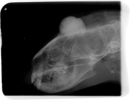
- A four-year-old ferret presented swelling of the top of the head of one month duration with a more rapid growing in the few last days. Examination and diagnostic imaging with radiography and computed tomography, revealed a bony mass of the temporal area of the skull. It measured between 1.2 and 1.5 centimeter in diameter.
Auteurs : Drs. Christophe Bulliot, Sophie Romain, Antoine Hidalgo, Elise Rattez, Alexandra Nicolier, Céline Levrier, Lucas Flenghi 10-07-2017
Service NAC – Centre Hospitalier Vétérinaire des Cordeliers,
29 avenue du Maréchal Joffre, 77100 Meaux.
E-mail : nac@chvcordeliers.com
Conférence Icare 2017 osteoma ferret
A case of calvarial osteoma and its surgical treatment in a ferret (mustela putorius furo)
It adhered to the frontal bone of about one centimeter in diameter. The portion of the frontal bone in contact with the encephalon presented no lesion. No abnormalities were revealed on the thoracic examination. Due to the rapid growth of the mass and the potential risk of encephalic compression, a surgical treatment was proposed and performed without previous biopsy.
Histologic diagnostic was calvarial osteoma. The ferret was still alive and in good health 14 months after surgery and a second computed tomography 8 months after surgery did not revealed any anomalies.
Osteomas are uncommon benign neoplasm of one bone. They arise mainly from the intermembranous bone of the skull and especially from the parietal bone, the zygomatic arch and occipital bone. Etiology is unknown. They are generally slow growing but the location and potential extent of the lesion can justify a poor prognosis.1-8 Two cases of ferrets presented with an osteoma of the parietal bone have been published in literature and have been euthanized because of poor body condition.9,10 To our knowledge, this is the first case report of successful surgical management of calvarial osteoma in a ferret.
References1. Dernell WS, Straw RC, Withrow SJ. Tumor of the skeletal system. In: Small Animal Clinical Oncology, 3rd ed. Philadelphia, USA, S.J. Withdraw and E.G. Mc Ewen W.B. Saunders; 2001:406-454. |