Le syndrome d’implantation septique (SIS) est un terme récent décrivant une endophtalmie septique associée à une rupture de la capsule cristallinienne avec une inflammation fibrinosuppurée centrée sur le cristallin et s’étendant en profondeur dans le cristallin.
Nous décrivons un tel cas survenu sur un chien de race husky sibérien, ayant malheureusement nécessité une énucléation.
Auteurs : Drs. P. Durieux et E. Papavasileiou 27-06-2018 Centre Hospitalier Vétérinaire des Cordeliers, 29-35 avenue du Maréchal Joffre, 77100 Meaux. E-mail : pdurieux@chvcordeliers.com Cet article a été publié dans : L’Essentiel (2018) 494 : p 26-29
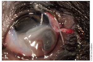
Une iridocèle associée à une déchirure de la capsule cristallinienne
La lacération cornéenne associée à une déchirure de la capsule antérieure du cristallin est fréquemment rencontrée chez nos animaux de compagnie après un traumatisme, notamment à la suite d’un coup de griffe par un chat.
Auteurs : Drs. P. Durieux et J. Taillandier 07-04-2018 Centre Hospitalier Vétérinaire des Cordeliers, 29-35 avenue du Maréchal Joffre, 77100 Meaux. E-mail : pdurieux@chvcordeliers.com Cet article a été publié dans : L’Essentiel (2018) 482 : p 27-30
Concentration en TNF-alpha dans l’humeur aqueuse de chiens sains ou atteints d’une pathologie intraoculaire : une étude pilote préliminaire
Summary
Objectives
To determine reference values of tumor necrosis factor-alpha (TNF-α) concentrations in the aqueous humor of control dogs. To show whether these values are significantly different from those obtained in dogs affected with intraocular pathology: acute anterior uveitis (AAU) or chronic primary angle closure glaucoma (PACG).
Auteurs : Drs. P. Durieuxa,∗, S. Etcheparebordea, D. Fritzb, S.G. Rosolenc, d27-01-2016
a Centre Hospitalier Vétérinaire des Cordeliers, 29 avenue du Maréchal Joffre, 77100 Meaux. b Companion animal laboratory (C.A.L), 1, rue Salomon-Rachi, 10000 Troyes, France c Inserm, U968, UPMC université Paris 06, UMR-S968, institut de la vision, 17, rue Moreau, 75012 Paris, France d Clinique vétérinaire Voltaire, 19, boulevard Voltaire, 92600 Asnières-sur-Seine, France E-mail : pdurieux@chvcordeliers.com Cet article a été publié dans : Journal français d’ophtalmologie (2015) 38, 288—294
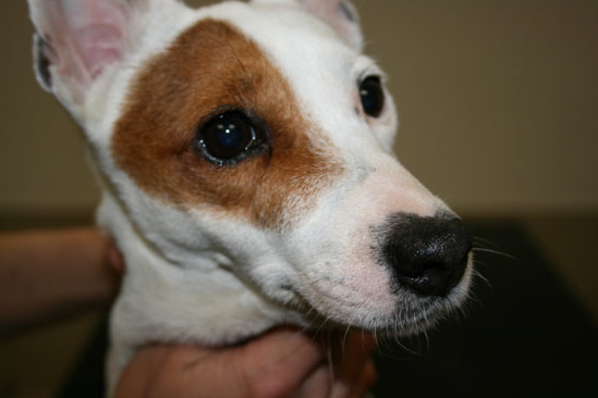
To describe a case of a nasolacrimal canaliculocele with intranasal extension in a dog. A 6-year-old neutered female Jack Russell Terrier was referred to the Centre Hospitalier Vétérinaire des Cordeliers for a slowly enlarging mass adjacent to the medial canthus of the right eye of 5 months duration.
Auteur :Drs. Philippe Durieux,* Stéphane Libermann,* Elise Rattez,* Marie Lagadic,† Yannick Ruel‡ and Thomas Chen§ 2015 E-mail : pdurieux@chvcordeliers.com Mots clefs : canaliculocele, CT, dog, endoscopy, marsupialisation, nasolacrimal Cet article a été publié dans : American College of Veterinary Ophthalmologists, Veterinary Ophthalmology, 18, 69–77
Purpose To evaluate the effects of levothyroxine (LTh) on the electroretinogram (ERG) of adult dogs.
Material and methods Binocular, full field photopic and scotopic ERGs were recorded from an anesthetized Maltese Bichon cross (MB), a Yorkshire Terrier (YT) and a Shetland Sheepdog (SS) affected with hypothyroidism and treated with a daily dose of LTh at 20μg/kg. The photopic ERGs were evoked to 12 different intensities ranging from 0.81 to–2.19 log cd.s/m2 and presented under photopic conditions in order to assess (from the derived luminance-response curves) Vmax and b : a amplitude ratio parameters. Photopic flicker ERGs were obtained at 30 Hz. The scotopic ERGs (intensity: –3.09 log cd.s/m2) were recorded while the retina was dark-adapting and after 32 min of dark adaptation.
Auteurs : Drs. Philippe Durieux,* F. Rigaudière,† J.-F. LeGargasson‡ and S. G. Rosolen‡§¶ *Clinique Vétérinaire, Meaux, France;†UDD Paris 7, France;‡INSERM U-592, UMR UPMC-Paris 6, Institut de la Vision, Paris, France;§CliniqueVétérinaire, Asnières, France;¶Fondation Ophtalmologique A. de Rothshild, Paris, France Centre Hospitalier Vétérinaire des Cordeliers, 29 avenue du Maréchal Joffre, 77100 Meaux. Mots clefs/Key Words : dog, electroretinogram, hypothyroidism, levothyroxine, photopic ERG, scotopic ERG E-mail : pdurieux@chvcordeliers.com Cet article a été publié dans : Veterinary Ophthalmology (2008) 11, 406–411
Correction de l’entropion inféro-latéral chez le chien par la technique chirurgicale décrite par Read et Broun en 2007 combinant la technique de Hotz Celsus et l’exérèse d’un coin palpébral latéral : étude rétrospective de 42 cas*
Correction of lateral lower lid entropion in dogs by the surgical technique described by Read and Broun in 2007 combining Hotz Celsus and lateral eyelid wedge resection: A retrospective study of 42 cases*